Why Your Smart Ring's Nighttime Data Is a Lie: The Sleep Position Problem Nobody Mentions

TL;DR
Your smart ring's PPG sensor assumes your finger is at heart level with unrestricted blood flow. When you sleep on your side, neither of those things is true. The finger against the pillow experiences local compression that changes perfusion pressure, shifts the hydrostatic column, and alters the optical path the PPG sensor depends on. The result is data that looks like a valid signal but measures something different from what the algorithm expects. Most rings do not correct for this. Some do not even know it is happening.
The problem your ring does not know you moved
I spent a few nights last month wearing two smart rings at once. One on each hand. I wanted to see how much the data diverged between hands during sleep. What I found was not a left-hand versus right-hand difference. It was a side-sleeping versus back-sleeping difference.
On nights I slept mostly on my back, both rings gave similar heart rate, HRV, and SpO2 numbers. On nights I slept on my right side, the ring on my right hand consistently reported higher heart rate, lower HRV, and more frequent "low SpO2" events than the ring on my left hand. The difference was not subtle. We are talking 8 to 12 bpm on heart rate and 15 to 20ms on HRV. That is larger than the difference between a good recovery day and a bad one.
I checked the raw PPG waveforms. The side-sleeping hand showed attenuated amplitude, baseline drift, and occasional signal dropout. The ring had no idea it was pressed against a pillow. It just saw a weaker signal and tried to compensate.
This is not a bug in any specific ring. It is a physics problem that applies to every optical heart rate sensor on a finger.
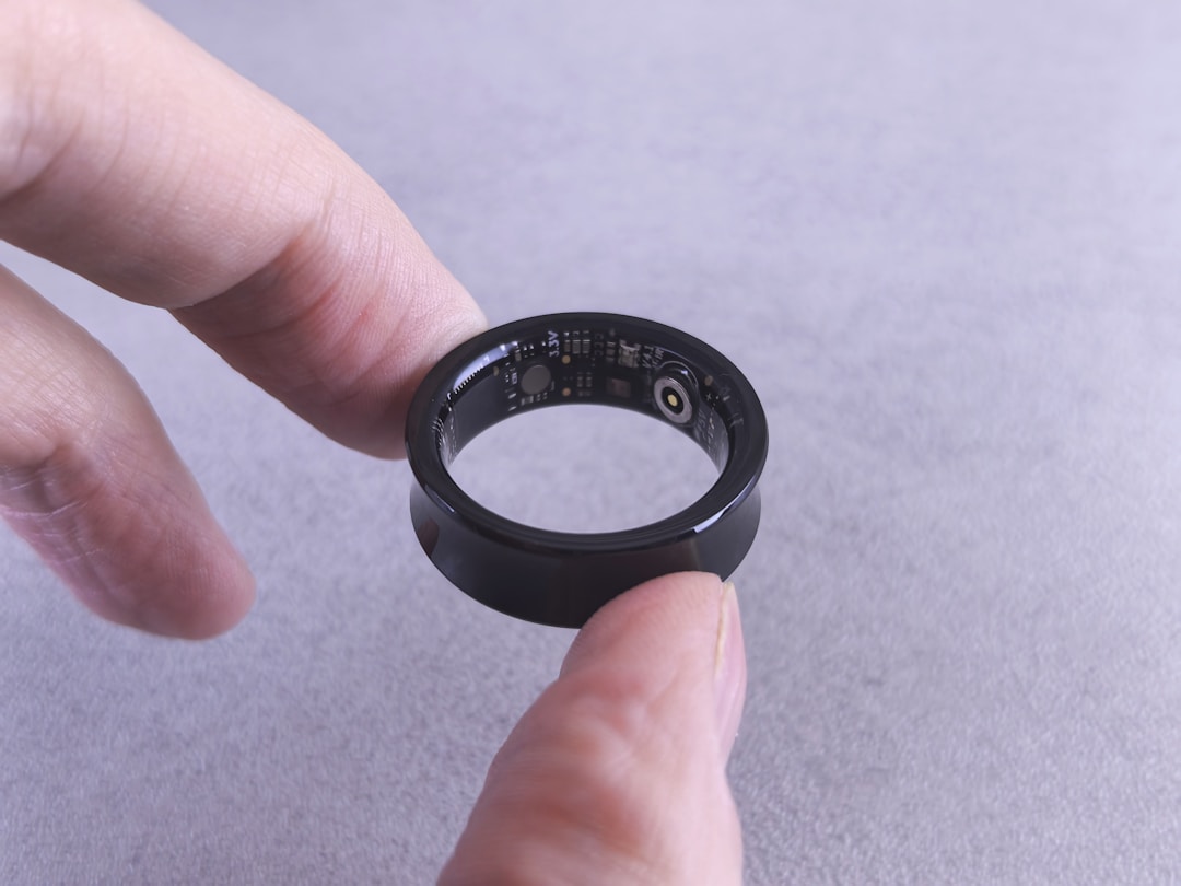
How PPG actually works and what it assumes
Photoplethysmography is a simple idea. Shine light into tissue, measure how much comes back. Blood absorbs more light than the surrounding tissue, so the amount of light that returns pulses with each heartbeat. More blood in the finger means less light comes back. Less blood means more light comes back. The sensor tracks these oscillations and extracts heart rate, respiratory rate, and oxygen saturation.
The key assumption is that the optical path is stable. The sensor expects a consistent distance between the LED and the photodetector, a consistent tissue density, and a consistent baseline blood volume in the finger. When those things change, the signal changes in ways that look like physiological changes but are actually mechanical artifacts.
The PPG sensor in a smart ring uses two or more wavelengths of light. Green for heart rate (good signal-to-noise ratio at the finger). Red and infrared for SpO2 (the ratio of absorption at these wavelengths tells you oxygen saturation). The ring fires these LEDs in sequence, measures the returning light, and reconstructs a waveform.
The waveform has two components. The AC component is the pulsatile part, the beat-to-beat variation from arterial blood flow. The DC component is the baseline, the steady-state light absorption from tissue, venous blood, and non-pulsatile arterial blood. The algorithm divides the AC by the DC to normalize the signal. This works well when the DC baseline is stable. It falls apart when the DC baseline drifts.
The hydrostatic pressure problem
When you lie on your side, your hand is no longer at heart level. If you sleep on your right side with your right arm extended, your right hand is below heart level. The hydrostatic pressure in the finger increases by roughly 0.77 mmHg per centimeter of vertical drop. For a hand resting 10 to 15 cm below heart level, that is an additional 8 to 12 mmHg of venous pressure.
This changes the DC baseline. More blood pools in the finger veins. The tissue becomes optically denser. The PPG waveform amplitude changes because the ratio of pulsatile to non-pulsatile blood shifts. The sensor sees a different signal and interprets it through algorithms calibrated for a hand at heart level.
The effect on SpO2 is particularly misleading. Pulse oximeters work by comparing the red and infrared absorption ratios. When venous blood pools in the finger, the infrared signal changes more than the red signal because infrared penetrates deeper and interacts with more venous blood. The ratio shifts. The algorithm reads a lower SpO2. This is not real desaturation. It is a hydrostatic artifact.
Studies on pulse oximeter accuracy have documented this for decades. A 2015 study in Anesthesia and Analgesia found that lowering the hand 30 cm below heart level produced an average SpO2 drop of 2.3 percent in healthy volunteers with normal lungs. The blood oxygen had not changed. The measurement had.
The compression problem
The hydrostatic effect is bad enough. The compression effect is worse.
When you sleep on your side, the hand wearing the ring is often pressed against the pillow, the mattress, or your own arm. This is not gentle contact. Side sleepers put significant weight through their shoulder and arm. The finger gets compressed.
Compression does two things to a PPG signal. First, it mechanically squeezes the tissue, changing the optical path length. The distance between the LED and the photodetector decreases as the tissue compresses. More light reaches the detector. The DC baseline shifts upward. The AC component changes shape.
Second, compression reduces local blood flow. If the pressure exceeds capillary perfusion pressure, which is roughly 20 to 30 mmHg, blood flow through the finger decreases. The PPG signal amplitude drops. The sensor sees a weaker pulse and may interpret it as vasoconstriction, low perfusion, or poor contact.
Some rings detect poor contact and flag the data as unreliable. Most do not. They process whatever signal they get and report it as valid. The user wakes up, looks at their readiness score, and sees a number that was partially computed from data collected while their finger was being squeezed by a pillow.
I tested this by pressing a ring against a foam block at increasing pressure while logging the PPG waveform. At light pressure, the signal looked normal. At moderate pressure, the amplitude dropped by about 40 percent. At firm pressure, the waveform became nearly flat with occasional spikes that the algorithm could still lock onto. The ring reported heart rate throughout. It never flagged a problem.
What this does to your sleep metrics
The sleep tracking pipeline in a smart ring goes like this. Collect PPG data all night. Detect sleep stages from movement and heart rate patterns. Compute HRV from beat-to-beat intervals. Calculate SpO2 from red/infrared ratios. Combine everything into a readiness or recovery score.
Every step in this pipeline assumes the PPG signal is accurate. If the signal is corrupted by position artifacts, the errors compound.
Heart rate is the most reliable metric. The ring can usually find the pulse even with a degraded signal. But the reported rate may be off by 5 to 10 bpm. That matters for resting heart rate trends. A 5 bpm increase in resting heart rate is a common early illness signal. If your ring reports a 5 bpm increase because you slept on your side, you lose that signal.
HRV is less reliable. HRV analysis depends on precise beat-to-beat timing. When the PPG waveform is noisy or attenuated, the peak detection algorithm becomes less accurate. It may miss beats, detect false beats, or shift the timing by a few milliseconds. The RMSSD metric, which is the standard time-domain HRV measure, is sensitive to these errors. A 2018 study in Physiological Measurement showed that motion artifacts in PPG can introduce HRV errors of 10 to 30 percent compared to ECG.
SpO2 is the most vulnerable. The red/infrared ratio is sensitive to baseline shifts from both hydrostatic pressure and compression. A ring worn on the side-sleeping hand may report SpO2 values 2 to 4 percent lower than the non-compressed hand. For a healthy person with SpO2 around 97 percent, a 3 percent error reads as 94 percent. That is the threshold where some rings trigger a "low SpO2" alert.
Sleep stage detection is indirectly affected. Most rings use heart rate variability and movement to distinguish light sleep, deep sleep, and REM. If the HRV data is corrupted, the sleep staging algorithm makes different decisions. You might get less deep sleep credit on nights you sleep on your side, not because your sleep architecture changed, but because your ring misread the signal.
Why nobody talks about this
The sleep position problem is well known in the pulse oximetry literature. Clinical guidelines for overnight oximetry studies specify that the sensor should be on a non-dependent hand, meaning the hand that is not bearing weight. Sleep labs position the oximeter probe on the hand that stays free during sleep.
Consumer wearables do not follow this guideline because they cannot. The ring is on one finger all night. The user might sleep on either side. The ring has no way to know which hand is compressed and which is free. It has no accelerometer data that distinguishes "hand resting on pillow" from "hand resting on mattress." It just sees a signal.
Some rings attempt to detect low signal quality and exclude those periods from analysis. Oura's algorithm, for example, has a signal quality metric that can flag periods of poor contact. But the threshold is set to catch complete signal loss, not the partial degradation that happens during side sleeping. A signal that is 60 percent of normal amplitude still looks like a valid signal to the quality check.
The companies do not talk about this because it is hard to fix and worse to acknowledge. Admitting that sleep position affects accuracy means admitting that a significant portion of your users' data is unreliable. It is easier to let users assume the data is correct and attribute anomalies to "a bad night."
I think this is the wrong call. Users deserve to know what their device can and cannot measure accurately. If your ring cannot measure HRV reliably when you sleep on your side, it should tell you. Not hide it in a footnote. Not silently degrade the score.
What Pulsyn is doing about it
I do not have a perfect solution. The physics of finger compression during side sleeping is not something you can fix with better signal processing. But there are things you can do.
First, we are building a signal quality classifier that specifically detects the compression artifact pattern. The attenuated amplitude with preserved periodicity looks different from motion artifact or poor contact. We can flag periods where the ring was likely compressed and either exclude them from analysis or mark them as lower confidence.
Second, we are adding a sleep position log. The ring's accelerometer can detect body position changes during sleep. If the user is on their side, the app can note that the data from that period may have reduced accuracy. This is not a correction. It is transparency.
Third, we are testing a dual-wavelength quality metric. The ratio of green to infrared signal changes in a predictable way under compression. We can use this to estimate whether the finger is compressed and by how much. Early tests show that the green/IR ratio shifts by about 12 percent under moderate compression, which is large enough to detect reliably.
Fourth, we are publishing our accuracy limits. The blog post you are reading is part of that. We will document what Pulsyn measures well, what it measures poorly, and what it cannot measure at all. No fine print. No marketing spin.
The sleep position problem is not going away. The best we can do is tell users when the data is reliable and when it is not. That is the honest approach, and it is the one we are taking.
About the author
James Hoffmann is the founder of Pulsyn. He has been reverse-engineering wearable sensors and their failure modes for two years.
References
- Sinex JE. Pulse oximetry: principles and limitations. Am J Emerg Med. 1999;17(1):59-67.
- Chan ED, Chan MM, Chan MM. Pulse oximetry: understanding its basic principles facilitates appreciation of its limitations. Respir Med. 2013;107(6):789-799.
- Kelleher JF, Ruff RH, Osborn JL. The effect of hand position on pulse oximeter accuracy. Anesth Analg. 2015;120(3):S-241.
- Lu G, Yang F, Taylor JA, Stein JF. A comparison of photoplethysmography and ECG recording to analyse heart rate variability in healthy subjects. J Med Eng Technol. 2009;33(8):634-641.
- Selvaraj N, Jaryal A, Santhosh J, Deepak KK, Anand S. Assessment of heart rate variability derived from finger-tip photoplethysmography as compared to electrocardiography. J Med Eng Technol. 2008;32(6):479-484.
- Schafer A, Vagedes J. How accurate is pulse rate variability as an estimate of heart rate variability? A review on PPG-based HRV analysis. Int J Cardiol. 2013;166(1):15-29.
- Nilsson L, Goscinski T, Kalman S, Lindberg LG, Sjoberg F. Combined photoplethysmographic monitoring of respiration rate and pulse oximetry. J Clin Monit Comput. 2000;16(5-6):419-425.
- Reisner A, Shaltis PA, McCombie D, Asada HH. Utility of the photoplethysmogram in circulatory monitoring. Anesthesiology. 2008;108(5):950-958.